
Resources
Frequently Asked Questions (FAQs)
about PaintScape™ direct visualization of the 3D genome with jebFISH™ technology
The PaintScape PLatform
What is PaintScape ?
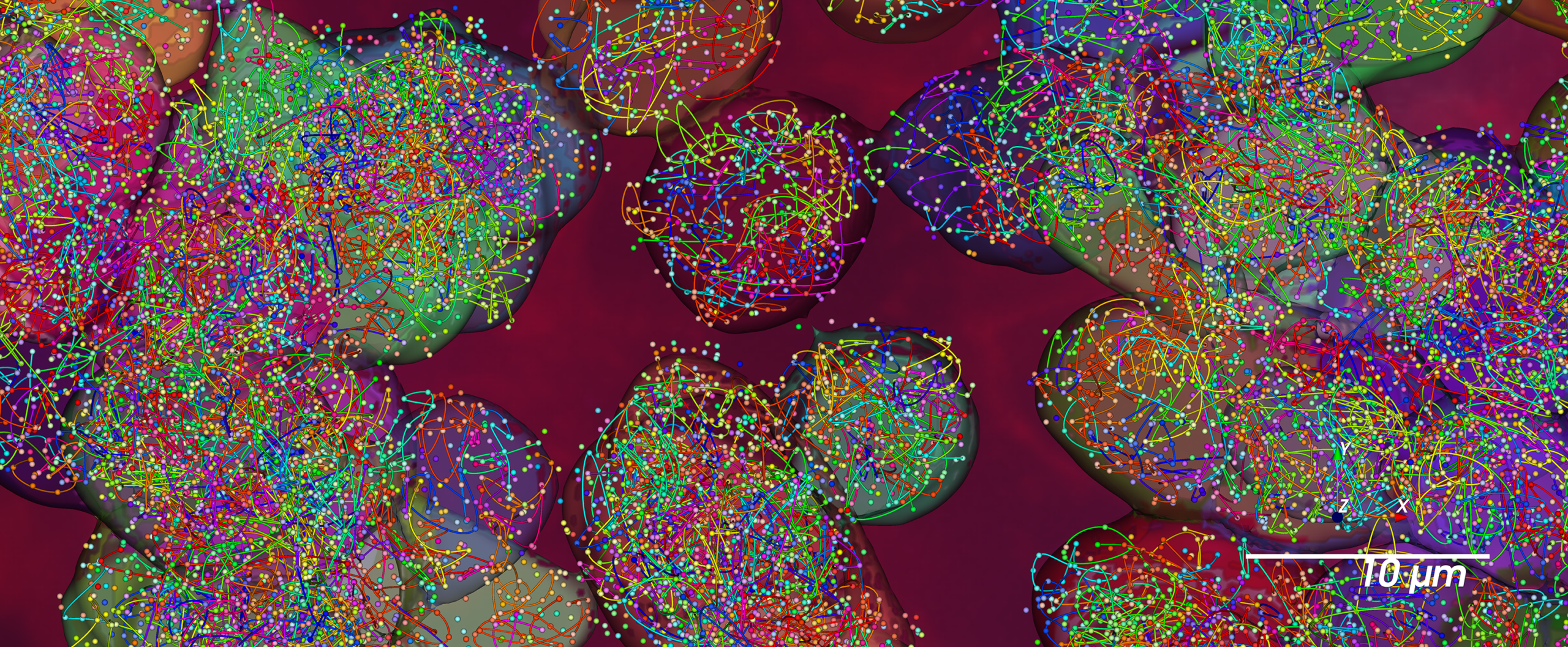
PaintScape (or officially the PaintScape™ System) is a groundbreaking spatial biology platform for directly visualizing genome organization (often called the 3D genome) in situ in single cells. The revolutionary PaintScape platform combines state-of-the-art optics, sophisticated bioinformatics, and a reagent system based on powerful jebFISH™ chemistry for direct in situ visualization of the 3D genome in individual cells at very high-resolution and very high multiplex.
The PaintScape platform makes it possible to directly see chromosomal structure—territories, conformation, and aberrations (including ploidy variation, copy number variation, segmented duplications, translocations, and ecDNA formation)—by enabling highly multiplexed “painting” of genomic regions of interest and visualizing chromosome traces. In addition, even finer levels of genome organization can be profiled such as A/B compartments, TAD (topologically associating domain) structure and boundary disruption, and nuclear position of targeted genomic loci.
With this capability, PaintScape direct visualization of the 3D genome elucidates mechanisms of structural genome dysregulation that correlate to disease and provides novel insights on genome disruption such as those found in cancer and in neurodegenerative disease.
The PaintScape platform includes robust, high-performance instruments; cutting-edge jebFISH-powered assay kits; sample carrier slide cartridges; and powerful bioinformatics analysis tools.
The components of the full PaintScape System are listed below:
•PaintScape™ Imaging Instrument
•PaintScape™ Sample Prep Instrument
•PaintScape™ jebFISH™ Assays
—ChromoPaint™ HuCL PanChromo MPX Kit
—OncoPaint™ HuCL Cancer Pathways Assay Kits
•PaintScape™ Accessories
—PaintScape™ Cell Line Chip
—PaintScape™ Tissue Chip (to be released)
•PaintScape™ Data Viewer
What is PaintScape genome painting?
PaintScape™ jebFISH™ genome painting is another name for PaintScape direct visualization of genome organization (the 3D genome) enabled by jebFISH protocols on the PaintScape Platform. In essence, PaintScape genome painting is the use of jebFISH chemistry and protocols on PaintScape instruments to directly visualize the 3D genome in single cells in situ in cell lines or fresh frozen tissue. The jebFISH method employs sophisticated barcoded oligonucleotide probes (LociPaint™ probes) to hybridize to target genomic loci regions in a highly multiplexed and efficient way and thus “paint” those genomic regions in situ.
What types of biological insights does PaintScape direct genome visualization reveal?
PaintScape direct visualization of the 3D genome elucidates structural genome mechanisms of dysregulation that correlate to disease such as cancer. With the PaintScape Platform, you can directly visualize and compare differences in ploidy, structural variations and chromosomal aberrations (e.g., translocations, focal amplifications, ecDNA), p-arm and q-arm variations, nuclear positioning, chromosome openness, or the proximity of multiple genomic regions of interest (not just two at a time) directly in situ in single cells.
You can then aggregate the data into sub- and whole cell populations and identify subsets of cells with particular genome organization profiles.
PaintScape reveals the following types of insights directly in situ in single cells:
•Ploidy aberrations, p-arm and q-arm variations for individual cells in situ in your sample
•Chromosome folding and positioning
•Structural variations such as focal amplifications and segmented copy number variation
•Translocations, ecDNA formation, and other chromosomal aberrations
•Nuclear position of genomic regions and A/B compartment analysis
•Genomic structural boundary aberrations; TAD (topologically associating domain) disruptions
•Degree of genomic region accessibility and openness
•Interaction proximities of multiple genomic loci regions simultaneously (not just binary interactions)
•and more…
(see featured PaintScape data for examples)
What are the components of the PaintScape Platform?
The PaintScape™ platform (officially the PaintScape System) includes robust, high-performance instruments, cutting-edge jebFISH-powered assay kits, sample carrier slide cartridges, and powerful bioinformatics analysis tools.
The components of the full PaintScape System are:
•PaintScape™ Imaging Instrument
•PaintScape™ Sample Prep Instrument
•PaintScape™ jebFISH™ Assays
—ChromoPaint™ HuCL PanChromo MPX Kit
—OncoPaint™ HuCL Cancer Pathways Assay Kits
•PaintScape™ Accessories
—PaintScape™ Cell Line Chip
—PaintScape™ Tissue Chip (to be released)
•PaintScape™ Data Viewer
What does the PaintScape Imaging Instrument do?
The PaintScape Imaging Instrument is a high-performance, robust imaging instrument for highly multiplexed, high-resolution jebFISH-powered direct visualization of the 3D genome in situ in cell lines or fresh frozen (FF) tissue. Designed with state-of-the-art, advanced optics and integrated fluidics; and the convenience of laboratory benchtop installation. Powerful data analysis tools are included, and various reports are available for analysis by single cell, sub-populations, or whole cell populations.
What does the PaintScape Sample Prep Instrument do?
The PaintScape™ Sample Prep Instrument provides streamlined sample preparation with automated fluidics and optimized protocols for highly multiplexed PaintScape jebFISH direct visualization of the 3D genome. The instrument was designed with researchers’ ease of use in mind and thus has simple touchscreen controls and walkaway automation. Moreover, by providing robust, automated sample preparation, the PaintScape Sample Prep Instrument reduces experimental variation from manual processes and enables researchers to focus on higher priority tasks.
Sample prep run parameters can be saved and re-used for later runs. The instrument is designed for use with PaintScape™ Cell Line Chips or PaintScape™ Tissue Chips (scheduled for future release) and enables sample prep of multiple PaintScape chips simultaneously.
Is PaintScape genome visualization a single cell method?
Yes — PaintScape™ analysis is a method to directly visualize the 3D genome in situ in single cells of human cell lines or fresh frozen tissue samples — resulting in genome painting directly in single cells and with spatial context. The jebFISH protocols are applied to the experimental sample directly on the slide; the PaintScape instruments then image and record data for each cell as well as across the slide. Data from individual single cells can be individually analyzed, pooled into specific cell sub-populations, or fully pooled for whole population analyses.
Thus, PaintScape can discover novel subpopulations with specific or unique genome architecture signatures and allows unprecedented, deeper study of selected or identified subpopulations.
Is PaintScape an in situ method?
Yes — PaintScape™ analysis using jebFISH™ technology is performed directly on cells in situ and enables visualization of the genome directly in individual single cells in situ in the experimental sample. Images and data are recorded for each cell as well as across the sample on the PaintScape platform instruments. Chromosome traces of each cell are visualized and imaged in situ — in position — and used for concomitant analyses including proximities of multi-loci genomic regions, inter-target distance heatmaps, and chromosome conformation and folding analyses.
Can PaintScape be done on interphase cells?
Yes — PaintScape™ jebFISH™ genome painting visualizes the 3D genome in situ directly in all cells in the sample regardless of which cell-cycle stage they are in. Since most cells in a given sample or cell population are in the interphase stage, most of the images and data recorded are of interphase cells. Moreover, PaintScape direct genome visualization is a very highly multiplexed method and thus enables painting and tracing of all chromosomes simultaneously and in very high resolution directly on individual cells in situ in the sample.
What sample types can PaintScape analysis be performed on?
PaintScape™ jebFISH™ genome painting can be performed on either cell lines or fresh frozen tissue. On cell lines, adherent cell lines work best. Currently, PaintScape jebFISH assay kits are commercially available for performing jebFISH-powered PaintScape 3D genome visualization on human cell lines. Commercially available PaintScape jebFISH assay kits for use on human fresh frozen (FF) tissue are in development and will be available at a later date. Posters presented at AGBT and AACR show results and novel insights from PaintScape jebFISH assays on FF human colorectal carcinoma tissue and on human glioblastoma tissue.
(see scientific posters for examples)
Is PaintScape genome painting a high-plex method?
Yes — PaintScape™ jebFISH™ genome painting is a very highly mulptiplexed method for directly visualizing the 3D genome in situ in single cells. jebFISH leverages multiplexed panels of LociPaint™ oligonucleotide probes that hybridize simultaneously; in fact, several hundreds to greater than a 1,000 genomic loci targets can be simultaneously visualized.
The first commercially available assay kit (ChromoPaint™ HuCL PanChromo MPX) contains a panel of 419 LociPaints that paint approximately evenly spaced genomic region targets across all chromosomes—including Chromosome Y.
The OncoPaint™ HuCL Cancer Pathways Assay Kits enable simultaneous painting of over 1,000 genomic loci targets that span known cancer pathway genes and regulatory regions from select cancer pathways. The kits are currently in development but available for pre-order.
Depending on the assay, each genomic region of interest is painted at approximately 5 to 15 genomic regions that span the targeted genomic loci (and often also the associated regulatory regions). By painting those genomic loci regions with multiple targets (i.e., like beads on a string), PaintScape jebFISH analysis provides chromatin folding/conformational detail and further structural genomic resolution of those regions—sometimes including TAD-level detail.
In addition, genomic targets that serve as A/B compartment analysis markers or other structural chromatin landmarks are also painted.
How many genome targets can be simultaneously visualized by PaintScape?
PaintScape™ jebFISH™ analysis is very highly multiplexed. Therefore, greater than a thousand genomic loci targets can be simultaneously detected and visualized.
Data presented in scientific posters at AGBT and AACR provide examples of studies where over 1,000 genomic loci targets were visualized simultaneously. In fact, PaintScape jebFISH genome painting has even visualized many thousands of loci targets in cells that developed severe aneuploidy (16X or more ploidy!).
What is so groundbreaking about PaintScape?
The groundbreaking aspect of PaintScape direct genome visualization is just that—the fact that you can directly visualize and see the genome in situ at high resolution and with high precision in individual single cells of cell lines or fresh frozen tissue. The power of directly seeing and visualizing genome organization in situ is profound.
Furthermore, the PaintScape Platform unleashes the breakthrough ability to trace chromosome homologs simultaneously directly in situ in individual cells. In other words, PaintScape traces every chromosome—in fact every homolog—directly in every cell across the sample.
Therefore with PaintScape, for the first time you can directly see chromosome traces (even in interphase cells) and see their conformation and relative positions within the nucleus in 3D space. And because PaintScape paints genomic regions of interest at both very high multiplex (>1,000 genomic loci targets) and at very high resolution, you can directly see chromosomal aberrations (e.g., translocations, ecDNA, focal amplifications, and p-arm/q-arm gain or loss), perform A/B compartment analysis, as well as see structural genome disruptions often with TAD-level detail.
Other methods for genome architecture study have been limited to inferred information from bulk analysis (e.g., in chromosome conformation capture (3C) methods like Hi-C) or to extremely low-plex, coarse resolution traditional DNA-FISH images. Instead, PaintScape empowers you to directly see genome architecture directly in situ in single cells at high resolution.
(see featured PaintScape data and scientific posters)
What type of detection signal is utilized in PaintScape?
PaintScape jebFISH™ analysis utilizes fluorescence imaging and fluorescently labeled oligonucleotide secondary reader probes (called jebReader™ Probes) to illuminate and detect the LociPaint™ oligonucleotide probes that have hybridized to the targeted genomic loci regions in situ.
What is a PaintScape image?
A PaintScape image is the key high-resolution, highly multiplexed starting image of in situ puncta from a PaintScape direct genome visualization run that provides the unprecedented ability to visualize chromosome traces and genome architecture directly in situ in single cells of a cell line or fresh frozen tissue sample.
(see featured PaintScape data and scientific posters)
A PaintScape image lets you see chromosomes, see chromosome traces (and thus their folding and conformation), see genome structural organization—in essence see the 3D genome directly in your cells in situ thus unleashing the power of direct visualization for revealing novel biological insights of structural genome mechanisms of health and disease.
In a PaintScape image, each targeted genomic region is illuminated, identified, and located in 3D space within each cell of the sample simultaneously with all targeted genomic regions. Therefore, PaintScape breaks revolutionary new ground and enables the powerful ability to directly see hundreds to 1,000+ genomic loci targets simultaneously in situ in single cells in the cell sample—and even as they exist tightly folded within the limited space of a cell nucleus.
PaintScape data analysis starts with this very high resolution and highly multiplexed image of the genome-wide targeted genomic regions in individual cells in situ across the cell sample.
(please see featured PaintScape data and scientific posters)
What kind of data analysis and insights does the PaintScape System provide?
As a high-precision, imaging-based genome analysis method, PaintScape unleashes the groundbreaking power of directly seeing and visualizing genome organization and tracing chromosomes directly in situ in individual single cells of cell line or fresh frozen tissue samples.
PaintScape data analysis thus starts with a very high-resolution and highly multiplexed PaintScape image of puncta that simultaneously illuminate genome-wide targeted genomic regions directly in individual single cells in situ across a cell sample. The imaged puncta are identified, located in 3D space, and then resolved into chromosomes traces.
Once captured in PaintScape images, the chromosome traces not only visualize each chromosome in situ in each cell, but also reveal their conformation, folding, and positions within the nucleus — thus also directly visualizing and unveiling genome insights in situ such as ploidy changes, copy number variation, p-arm/q-arm variations, focal amplifications, ecDNA formation, translocations, inter-target distances, multi-loci proximities, chromatin compaction, A/B compartment segregation, TAD boundary disruption, and nuclear position. (see featured PaintScape data and scientific posters for examples)
Importantly, PaintScape genome imaging lets you not only directly visualize these phenomena for each single cell and each chromosome traced but also for pooled cell populations and sub-populations. Thus, PaintScape can identify subsets of cells with specific genome architecture profiles for deeper analysis or to reveal biological trends among them.
PaintScape images and data analysis outputs include:
• High-resolution, highly multiplexed PaintScape image of genome targets painted in situ in cell sample (cell lines or fresh frozen tissue) with cell segmentation layer
• Image overlays of chromosome traces visualized in situ (with ability to view one, a select few, or all chromosomes simultanesouly) with capability to zoom into chromosome traces for specific individual single cells and specific chromosomes in specific single cells
• Chromosome ploidy enumeration – calculated for individual single cells and also pooled to sub-populations or whole populations
• p-arm/q-arm enumeration and folding (viewable by individual homologs in individual single cells)
• Genomic target copy number enumeration – calculated for individual single cells and also pooled to sub-populations or whole populations
• Inter-target distance heatmaps calculated from aggregated spatial positions of all painted genomic targets from each imaged cell across the cell sample
• Various other plots, graphs, and heatmaps of measurements (calculated from PaintScape imaging data and chromosome traces) that return data on chromosome compaction/openness; A/B compartment segregation, TAD-level boundary disruptions, and many more features.
(see scientific posters and PaintScape featured data for examples)
What is the difference between PaintScape and traditional DNA FISH?
The main difference between PaintScape™ direct genome visualization and DNA FISH is the tremendous and fundamentally important difference in plex and resolution with which chromosomal DNA is and painted and imaged. PaintScape can target and image hundreds to 1,000+ genomic loci targets simultaneously while traditional DNA FISH usually only detects a 1-2 targets at a time.
Furthermore, the extremely high-plex nature of PaintScape genome painting means that PaintScape is able to trace the chromosomes and visualize their folding and conformation; not just return a few coarse spots to indicate the presences of a target. The power of chromosome tracing and seeing the chromosome conformations in situ unlocks whole new sets of information regarding the genome organization of the assayed cells.
While PaintScape direct genome visualization uses fluorescence in situ hybridization (FISH) principles, it utilizes an advanced, highly multiplexed version of FISH and additionally leverages proprietary, sophisticated jebSmart™ multiplex optical barcode technology in jebFISH™-chemistry-powered protocols on high-performance PaintScape instruments — thus, providing a groundbreaking high-plex, high-resolution way to directly visualize genome architecture in individual single cells in situ. Traditional DNA FISH, on the other hand, detects only a couple of coarse genomic regions at a time and often with only lower resolution imaging methods (traditional microscopy).
Moreover, PaintScape™ in situ genome analysis leverages panels of multiplexed, barcoded LociPaint™ oligonucleotide probes instead of the longer DNA probes commonly used in traditional DNA FISH. This additional key difference means that in PaintScape jebFISH analysis, the targeted regions of the genome are painted not only in a very highly multiplexed way but also with much finer detail and resolution than is possible with the larger DNA probes used in traditional DNA FISH.
Thus, PaintScape not only enables direct visualization of chromosome traces but also the ability to directly visualize and see select genomic regions of interest even with TAD-level detail. Therefore, PaintScape jebFISH visualizes the in situ 3D chromosomal structure of the genomic regions painted and also provides insights into chromatin openness and multi-loci spatial proximities.
(see scientific posters and featured PaintScape data for examples)
How is PaintScape genome visualization different from Hi-C or other chromosome conformation capture (3C) methods?
Both PaintScape direct genome visualization and the various chromosome conformation capture (3C) analysis methods (such as Hi-C) provide ways to study genome-wide genome organization. However, PaintScape, as an inherently high-resolution, in situ imaging and direct visualization method, provides 3D genome architecture insights directly in situ for individual single cells in a sample, and moreover, even directly visualizes and traces each individual chromosome in each single cell.
In contrast, Hi-C and other 3C methods are generally performed on bulk samples, and therefore, usually cannot return data for individual single cells nor can they provide direct information on specific chromosomes. Secondly and of important note, Hi-C and similar methods only provide inferred information on proximities between only binary regions of genomic DNA because those methods depend on detection of artificially crosslinked fragments of chromatin that are run through various chemistries (crosslinking, ligation, NGS) and require detection via next-gen sequencing reads.
Thus to recap, PaintScape jebFISH genome analysis directly visualizes genome architecture (the 3D genome) and directly traces each chromsome in situ in single cells whereas Hi-C and other 3C methods instead only return inferred, aggregate information from sequencing various detected species of artificially crosslinked stretches of DNA. Thus, while Hi-C and similar methods provide orthogonal, complementary information, PaintScape jebFISH technology breaks new ground and allows researchers to now investigate genome organization directly in situ in individual single cells and directly trace individual chromosome homologs in situ—providing deeper insights with in situ single cell detail and spatial context.
In addition with PaintScape jebFISH analysis, proximities of multiple genomic loci regions are revealed simultaneously (not just two at a time as in Hi-C). Also, because of the single cell detail, individual cells or cell sub-populations with specific genome architecture profiles can be identified and further investigated.
Lastly, PaintScape direct genome visualization can be performed in situ, not only on cell lines, but also on fresh frozen (FF) tissue samples. While a commercially available tissue assay is still in development, recent posters have demonstrated the utility and useful insights revealed from PaintScape jebFISH performed on FF tissue samples. While some Hi-C methods for use on tissue samples are starting to emerge, those methods remain extremely challenging, and the majority of Hi-C analysis is still currently constrained to bulk samples whether pooled from tissue or cell culture.
What assays are available for PaintScape?
Currently there are two types of highly multiplexed, genome-wide assays available for PaintScape:
•the ChromoPaint™ HuCL PanChromo MPX Assay — a survey assay for general genome conformation and architecture
•the OncoPaint™ HuCL Cancer Pathways Assay Kits — deeper structural analysis of select cancer pathway genes with genome-wide context of genome architecture
The initially released assay kits are designed for use on human cell lines; however, assay kits for use on human fresh frozen (FF) tissue will be released in the future and data/results from PaintScape genome visualization on FF tissue has been presented in recent scientific posters.
(also see the next question for information on the key differences between these assays)
What’s the difference between the ChromoPaint and OncoPaint kits?
There are two types of highly multiplexed, genome-wide assays currently available for PaintScape:
•the ChromoPaint™ HuCL PanChromo MPX Assay
•the OncoPaint™ HuCL Cancer Pathways Assay Kits
The ChromoPaint™ HuCL PanChromo MPX Assay Kit is designed as a general survey mode assay to assess basic genome conformation; the OncoPaint™ HuCL Cancer Pathways Assay Kits, on the other hand, enable additional, deeper, more detailed structural analysis of genomic regions that span select cancer pathway genes while also providing genome-wide context of chromosomal architecture.
The ChromoPaint Kit includes a panel of LociPaint™ probes for 419 genomic loci targets that are approximately, evenly spaced across all chromosomes; ChromoPaint is thus ideal for genome-wide target copy number assessment, chromosome ploidy enumeration, and detection of large-scale structural variations such as chromosome arm gain/loss, translocations, segmented copy number changes, ecDNA formation, folding, compaction, and nuclear position.
The OncoPaint Cancer Pathways Assay kits paint the genome with the very high multiplex of 1000+ genomic loci targets and provide the option to choose from select cancer pathway gene modules (initially from the following cancer pathways: cell cycle & apoptosis; DNA repair; transcriptional regulation; and chromatin modification) to pair with the core modules of chromatin structure and chromosome ploidy/compartment targets to perform a PaintScape jebFISH genome painting that not only paints all the chromosomes, but also provides finer structural detail in the genomic regions of the targeted pathway genes. OncoPaint Cancer Pathway Assays can thus paint greater than 1,000 genomic loci targets. Each targeted pathway gene is painted at approximately 15 genomic loci and thus can provide TAD-level detail.
The OncoPaint Assays are therefore ideal for investigating finer genome structural changes along cancer pathway genes; for example, revealing focal amplifications, translocations, openness, A/B compartments, TAD structure, and ecDNA formation in those genomic regions.
Both assay kits include reagents for sample preparation as well as imaging and readout of the hybridized LociPaint probe panels.
jebFISH Technology
What is jebFISH?
PaintScape™ jebFISH™ technology is a revolutionary spatial biology method for directly visualizing genome organization (the 3D genome) in single cells in situ on the PaintScape Platform — and thus adds a whole new type of omic analysis (that of genome architecture) for translational and discovery research in human health and disease.
In essence, PaintScape jebFISH technology is a set of highly multiplexed, efficient chemistries for in situ visualization of the 3D genome directly in single cells (in cell lines or fresh frozen tissue) and leverages both advanced, high-plex FISH (fluorescence in situ hybridization) protocols as well as sophisticated proprietary multiplexed optical barcodes called jebSmart™ barcoding.
PaintScape jebFISH chemistry enables simultaneous painting of greater than a 1,000 genomic loci regions in a way not possible before and thus the direct visualization and tracing of chromosomes in situ. The jebFISH protocols are performed on the PaintScape platform instruments which include the specially designed PaintScape™ Sample Prep Instrument and the PaintScape™ Imaging Instrument with state-of-the-art optics and bioinformatics. The combination of the proprietary jebSmart barcoding and the best-in-class optics of the PaintScape Imaging Instrument allow precision in target loci identification and localization, and thus, the unprecedented ability to visualize chromosome architecture directly in individual single cell nuclei in situ.
Thus with these capabilities, Paintscape jebFISH genome visualization makes it possible to directly see chromosomal structure—territories, conformation, and aberrations (including copy number variation, segmented duplications, translocations, ecDNA formation, and chromosome openness)—by enabling highly multiplexed “painting” of genomic regions of interest and visualizing chromosome traces with high resolution.
(see the jebFISH Workflow page for an overview diagram)
What is jebFISH genome painting?
PaintScape™ jebFISH™ genome painting is another name for PaintScape direct visualization of genome organization (the 3D genome) enabled by jebFISH protocols on the PaintScape Platform. In essence, jebFISH genome painting is the use of jebFISH chemistry and protocols on PaintScape instruments to directly visualize the 3D genome in single cells in situ in cell lines or fresh frozen tissue. The jebFISH method employs sophisticated proprietary jebSmart™ barcoded oligonucleotide probes (LociPaint™ probes) to hybridize to target genomic loci regions in a highly multiplexed and efficient way and thus “paint” those genomic regions in situ.
What are LociPaint probes?
LociPaint™ probes are specially designed oligonucleotide probes used in PaintScape jebFISH chemistry for direct visualization of the 3D genome. Designed using advanced bioinformatics and sophisticated barcoding, LociPaint probes hybridize to targeted genomic DNA regions of interest in a highly multiplexed way—thus enabling simultaneous and efficient painting of genomic loci targets at high-plex and with high resolution.
LociPaint probes are designed and used in sets. During a PaintScape jebFISH genome painting, multiple LociPaint probes hybridize to each targeted region—thus ensuring robust detection of that region during the imaging phase.
(see the jebFISH Workflow page for diagram)
What are jebReader probes?
jebReader™ probes are fluorescently labeled oligonucleotide probes that serve as secondary readout probes. By hybridizing to the LociPaint probes, they detect, illuminate, and identify the hybridized LociPaints. During the imaging phase of PaintScape jebFISH painting, each LociPaint probe is illuminated by multiple jebReader probes. The fluorescent signal from the jebReaders appear as puncta that are then located in 3D space and identified by decoding the fluorescent barcode signals.
(see the jebFISH Workflow page for diagram)
The 3D Genome
What is the 3D genome?
The 3D Genome (also referred to as genome organization or genome architecture) is the organization and structure of the genome —in essence all of a cell’s genomic DNA coiled and folded into chromatin and chromosomes and how it is folded and structured within a cell’s nucleus. It is now known that this “genome organization”—the 3D genome—plays an important role in cell programming and function.
Although all cells theoretically have a full set of “genomic blueprints” (a full genome) to carry out their cellular functions, the structural organization of the genome (not just its linear sequence) within each individual cell has impact on the regulation of those cellular programs and their dysfunction when things go awry. Moreover, chromatin folding patterns can reveal key insights. The dynamic folding and unfolding of sections of chromatin (especially those containing regulatory elements like enhancers and promoters) within each cell are critical to its ability to regulate cell division, gene expression, and regulatory pathways.
Structural genomic disruptions, chromatin architecture and accessibility, and chromosomal aberrations such as aneuploidy, focal amplifications, translocations, and formation of ecDNA are known to be key biological mechanisms in disease. Thus, research on genome architecture and the 3D genome has gained increased interest and remains an important area of research.
(see About the 3D Genome for diagrams and more info)
Why study the 3D genome (genome organization)?
While the human genome has been fully sequenced, the organization of the genome remains less studied and yet plays a key role in cellular programming and function. Although all cells theoretically have a full set of “genomic blueprints” (a full genome) to carry out its cellular functions, the organization and structure of the genome (not just its linear sequence) within each individual cell has impact on the regulation of those cellular programs and their dysfunction when things go awry.
Chromatin folding patterns, in fact, can reveal key insights. Only 2% of the genome carries protein-coding DNA; the other 98% includes critical regulatory regions such as enhancers and promoters. Thus, the dynamic folding and unfolding of sections of chromatin (especially those containing regulatory elements) within each cell is critical to its ability to regulate cell division, gene expression, and regulatory pathways. Furthermore, revealing the location, openness, and proximities of genomic loci regions relative to each other can reveal key biological understanding about structural genome mechanisms of disease.
Additionally, the very dynamic nature of chromatin folding means that not only are there differences in patterns of conformation and genome organization for different cells from different tissues or from different cell types within the same tissue, but that there are even differences and uniqueness of the genome architecture (the 3D genome) in individual single cells of the same cell type — even those grown in a single cell culture. In other words, each single cell in a given culture can carry a unique chromatin architecture and thus unique genome organization. PaintScape jebFISH genome painting provides the unprecedented ability to identify those unique 3D geome profiles in individual single cells in situ as well as unveil the patterns and frequencies of particular genome organization profiles in specific subsets of cells.
Why is it helpful to have single-cell specific data when studying the 3D genome?
The genome organization (or 3D genome) of a given individual single cell plays an important role in its cell programming and function. The structural organization of the genome within each individual cell has impact on the regulation of those cellular programs and their dysfunction when things go awry. In fact, chromatin folding patterns can critically impact an individual cell’s unique biology. In other words, the unique dynamic folding of sections of chromatin (especially those containing regulatory elements) within each cell is critical to its ability to regulate cell division, gene expression, and regulatory pathways.
The very dynamic nature of chromatin folding means that not only are there differences in patterns of chromatin conformation and genome organization for different cells from different tissues (or from different cell types within the same tissue or sample), but that there are also even differences and uniqueness of the genome architecture in individual single cells of the same cell type — even those grown in a single cell culture. In essence, each single cell in a given culture can hold a unique chromatin architecture and thus unique genome organization. PaintScape jebFISH genome painting provides the unprecedented ability to identify those unique 3D geome profiles in individual single cells in situ as well as unveil the patterns and frequencies of particular genome organization profiles in specific subsets of cells.
The unique genome profile of any individual cell may sometimes be attributed to be inocuous random variation and a certain degree of cell-to-cell heterogeneity may perhaps be expected in any population of cells — especially given the dynamic nature of chromatin folding. However, when the deviation results in an impactful aberrant genome architecture, it is crucial to be able to detect and identify those unique but significant individual variants. For example, when regulatory regions are folded in novel positions and conformations that lead to novel juxtapositions between chromatin segments and genomic regions, the resultant genome structural change can lead to concomitant instances of aberrant gene expression or gene dysregulation.
Identification of single cells that serve as initiators of such impactful clonal heterogeneity within a cell population can provide critical insights to disease initiation mechanisms and clues to how the heterogeneity pervades a clonal cell population.
Why is it hard to study the 3D genome?
Until now, it has been very challenging to directly investigate genome organization (including components such as chromatin and chromosomes) within the extremely confined space of a cell nucleus (often only 5–8 µm in diameter) and given that chromatin is so tightly packed within it.
It takes approximately 1.8m of DNA to hold a full human genome, and yet all that DNA needs to be packed and fit inside the tiny cell nucleus (in essence, a space about 6 orders of magnitude smaller!). To make that possible, there is important order and structure to how it is packed and also in how the resulting chromatin architecture and chromosomes are positioned and folded. Thus, to investigate the genome architecture, in situ, it takes innovative and efficient chemistry, protocols, and high-performance instrumentation. The jebFISH-powered PaintScape Platform provides that groundbreaking technology to breakthrough those challenges and enable direct visualization of genome organization (the 3D genome) in situ single cells and thus open up new scientific frontiers that can reveal novel discovery in genome biology for health and disease.
Examples of BIOLOGICAL INSIGHTS FROM PaintScape Direct 3D Genome Visualization
In Situ Chromosome Traces in Single Cells

(all chromosome view of a single nucleus)
Visualization of
Translocations

(Only Chr 3 and 6 displayed for clarity).
Structural Genome Insights across Cell Populations

PaintScape — a revolutionary spatial biology platform for visualizing the 3D genome directly in single cells
The PaintScape™ System is a high-resolution, highly multiplexed spatial biology genome analysis platform for direct visualization of the 3D genome in situ in single cells and combines state-of-the-art optics, sophisticated bioinformatics, and a reagent system based on powerful jebFISH™ chemistry.
PaintScape Inquiry


Talk to our expert team
Request a meeting to learn how PaintScape can help you discover novel genome insights.